
Dead Arm Syndrome is a perplexing condition that affects individuals engaged in repetitive shoulder movements causing sudden, acute pain that temporarily weakens or impairs the arm.

It often creeps up on athletes, especially baseball pitchers, who demand the utmost from their shoulders, only to find themselves grappling with sharp discomfort and a concerning loss of function.

In order to use your arm with minimal to no pain, it involves identifying the causes and symptoms, and seeking interventions that can alleviate discomfort and restore function.

A crucial step in this journey is having an informed conversation with a doctor or specialist with expertise in sports injuries.

Sports, tennis and arm pain on court after training, game or match outdoors. Healthcare, tennis player and injured black man or athlete with muscle pain or inflammation after exercise or workout

Introduction to Dead Arm Syndrome

Dead arm syndrome is a condition that can cause sudden, sharp arm pain. Athletes – such as football players or baseball players – may notice it because they can no longer throw.

An effective approach to treating dead arm syndrome not only relieves the immediate discomfort but also paves the way for ongoing health and optimal athletic performance.

Definition and Overview

Imagine if you were once able to use your throwing arm without pain and suddenly you can’t throw because your limb feels like it’s not your own. This confusing sensation is often experienced by those with dead arm syndrome, where the arm’s function is impaired without apparent reason, leading to discomfort and a hindered ability to perform certain physical activities.

Consulting with a physician upon noticing these symptoms is crucial, as dead arm syndrome may stem from a variety of underlying conditions, ranging from nerve compression, tissue inflammation or injury. Accurate diagnosis is essential, and a specialist’s expertise can pinpoint the root cause and provide an effective treatment plan.

Importance in Orthopedics

Within orthopedics, dead arm syndrome garners significant attention due to its potential to sideline athletes. It’s important to identify the issues so that individuals can reengage with their sports swiftly and, most importantly, safely.

A good understanding of dead arm syndrome allows doctors to deliver care that extends beyond managing symptoms. Treatments may include preventative strategies, an understanding of sports biomechanics, and tailored treatments, all working collaboratively to rehabilitate and fortify the arm against future injury.

Symptoms of Dead Arm Syndrome

Recognizing the symptoms of dead arm syndrome is key in tackling the condition head-on.

  • Pain and weakness:
    • Significant discomfort or pain in the shoulder, especially when throwing, accompanied by noticeable weakness in the upper arm.
  • Numbness and tingling:
    • A sensation of pins and needles or numbness radiating from the shoulder down the arm.
  • Reduced throwing ability:
    • Difficulty throwing with the same force and control as before, often experiencing a significant drop in throwing speed.
  • Shoulder instability:
    • Clicking or popping sounds in the shoulder joint, sometimes accompanied by a feeling of the shoulder partially dislocating.
  • Fatigue in the arm:
    • Feeling of tiredness and exhaustion in the affected arm, especially during repetitive movements.

As you better understand the signs and symptoms of dead arm syndrome, keep in mind that the characteristics of your discomfort and functional impairment offer critical clues in formulating an effective treatment plan.

Conclusion and Takeaways

Arm health and function depend on the timely recognition of symptoms, an accurate diagnosis, and a well-defined treatment plan. Noticing the signs and symptoms early, will help prevent prolonged discomfort and performance setbacks

It’s important you talk with your doctor to understand the underlying causes of your symptoms so that you can get treatment as soon as possible. Staying informed and supported through recovery is going to help with peak arm function and overall musculoskeletal health.

The physicians and specialists at Southeast Orthopedic Specialists are experts in all aspects of orthopedic and spine care. No matter the type of treatment you require, the team at Southeast Orthopedic Specialists is ready to serve you at one of their six locations across Duval, St. Johns and Clay counties. If you find yourself experiencing any type of pain related to an orthopedic or spine condition, call Southeast Orthopedic Specialists at 904.634.0640 or schedule an appointment online

(Photo Credit – Adobe)


Understanding Dead Arm Syndrome: Symptoms, Causes, and Treatment

Sports, tennis and arm pain on court after training, game or match outdoors. Healthcare, tennis player and injured black man or athlete with muscle pain or inflammation after exercise or workout

Dead Arm Syndrome is a perplexing condition that affects individuals engaged in repetitive shoulder movements causing sudden, acute pain that temporarily weakens or impairs the arm.

It often creeps up on athletes, especially baseball pitchers, who demand the utmost from their shoulders, only to find themselves grappling with sharp discomfort and a concerning loss of function.

In order to use your arm with minimal to no pain, it involves identifying the causes and symptoms, and seeking interventions that can alleviate discomfort and restore function.

A crucial step in this journey is having an informed conversation with a doctor or specialist with expertise in sports injuries.

Sports, tennis and arm pain on court after training, game or match outdoors. Healthcare, tennis player and injured black man or athlete with muscle pain or inflammation after exercise or workout

Introduction to Dead Arm Syndrome

Dead arm syndrome is a condition that can cause sudden, sharp arm pain. Athletes – such as football players or baseball players – may notice it because they can no longer throw.

An effective approach to treating dead arm syndrome not only relieves the immediate discomfort but also paves the way for ongoing health and optimal athletic performance.

Definition and Overview

Imagine if you were once able to use your throwing arm without pain and suddenly you can’t throw because your limb feels like it’s not your own. This confusing sensation is often experienced by those with dead arm syndrome, where the arm’s function is impaired without apparent reason, leading to discomfort and a hindered ability to perform certain physical activities.

Consulting with a physician upon noticing these symptoms is crucial, as dead arm syndrome may stem from a variety of underlying conditions, ranging from nerve compression, tissue inflammation or injury. Accurate diagnosis is essential, and a specialist’s expertise can pinpoint the root cause and provide an effective treatment plan.

Importance in Orthopedics

Within orthopedics, dead arm syndrome garners significant attention due to its potential to sideline athletes. It’s important to identify the issues so that individuals can reengage with their sports swiftly and, most importantly, safely.

A good understanding of dead arm syndrome allows doctors to deliver care that extends beyond managing symptoms. Treatments may include preventative strategies, an understanding of sports biomechanics, and tailored treatments, all working collaboratively to rehabilitate and fortify the arm against future injury.

Symptoms of Dead Arm Syndrome

Recognizing the symptoms of dead arm syndrome is key in tackling the condition head-on.

  • Pain and weakness:
    • Significant discomfort or pain in the shoulder, especially when throwing, accompanied by noticeable weakness in the upper arm.
  • Numbness and tingling:
    • A sensation of pins and needles or numbness radiating from the shoulder down the arm.
  • Reduced throwing ability:
    • Difficulty throwing with the same force and control as before, often experiencing a significant drop in throwing speed.
  • Shoulder instability:
    • Clicking or popping sounds in the shoulder joint, sometimes accompanied by a feeling of the shoulder partially dislocating.
  • Fatigue in the arm:
    • Feeling of tiredness and exhaustion in the affected arm, especially during repetitive movements.

As you better understand the signs and symptoms of dead arm syndrome, keep in mind that the characteristics of your discomfort and functional impairment offer critical clues in formulating an effective treatment plan.

Conclusion and Takeaways

Arm health and function depend on the timely recognition of symptoms, an accurate diagnosis, and a well-defined treatment plan. Noticing the signs and symptoms early, will help prevent prolonged discomfort and performance setbacks

It’s important you talk with your doctor to understand the underlying causes of your symptoms so that you can get treatment as soon as possible. Staying informed and supported through recovery is going to help with peak arm function and overall musculoskeletal health.

The physicians and specialists at Southeast Orthopedic Specialists are experts in all aspects of orthopedic and spine care. No matter the type of treatment you require, the team at Southeast Orthopedic Specialists is ready to serve you at one of their six locations across Duval, St. Johns and Clay counties. If you find yourself experiencing any type of pain related to an orthopedic or spine condition, call Southeast Orthopedic Specialists at 904.634.0640 or schedule an appointment online

(Photo Credit – Adobe)


Understanding Knee Pain and How Southeast Orthopedic Specialists Can Help

Knee Pain Treatment Specialist Exam's Orthopedic Patient

Knee pain is a common issue that affects individuals of all ages. Whether it’s caused by injury, arthritis, or overuse, knee pain can significantly impact your daily life and activities. In St. Augustine, many individuals are seeking expert treatment to alleviate their knee pain and improve their quality of life. 

Knee Pain Treatment Specialist Exam's Orthopedic Patient

The good news is that Southeast Orthopedic Specialists, with its specialized team of knee pain specialists, offers a variety of treatment options to help you keep life in motion.

What Causes Knee Pain?

Knee pain can result from several factors, including injury, chronic conditions like arthritis, or wear and tear from age or sports activities. Some of the most common causes include:

  • Arthritis: Both osteoarthritis and rheumatoid arthritis can lead to knee pain and stiffness, making movement difficult.
  • Sports Injuries: ACL tears, meniscus injuries, and patellar tendinitis are common among athletes.
  • Overuse: Repetitive movements or prolonged activity can lead to knee pain, especially in those who engage in physical labor or sports.
  • Bursitis: Inflammation of the small fluid-filled sacs in the knee can cause swelling and discomfort.

Knee Pain Treatment Options at Southeast Orthopedic Specialists

At Southeast Orthopedic Specialists, we understand that knee pain can be debilitating, which is why we offer comprehensive, customized treatment plans tailored to your unique needs. Our team of knee pain specialists in St. Augustine is dedicated to providing advanced care that focuses on long-term relief and recovery.

Non-Surgical Knee Pain Treatments
We offer a variety of non-invasive treatments for knee pain, including:

  • Physical Therapy for Knee Pain: Our physical therapists work with you to develop personalized exercise programs designed to strengthen the muscles around the knee and improve mobility.
  • PRP Therapy for Knee Pain: Platelet-rich plasma (PRP) therapy utilizes your body’s own healing properties to promote tissue repair and reduce inflammation, especially in cases of arthritis or soft tissue injuries.
  • Knee Braces: Custom knee braces provide support and stability, helping alleviate pain and protect the joint during daily activities.
  • Pain Management Solutions: From injections to targeted therapies, we offer treatments that focus on reducing inflammation and managing chronic knee pain.

Surgical Options
For more severe cases, our orthopedic knee surgeons are skilled in minimally invasive procedures such as knee arthroscopy and joint replacement, ensuring a swift recovery with optimal results.

Why Choose Southeast Orthopedic Specialists for Knee Pain Relief in St. Augustine?

Southeast Orthopedic Specialists stands out as a leading provider of orthopedic care in St. Augustine, offering specialized knee pain treatment that helps patients get back to their active lifestyles. Our comprehensive approach includes:

  • Expert Specialists: We have some of the top knee specialists in St. Augustine, with extensive experience in diagnosing and treating all forms of knee pain.
  • Cutting-Edge Treatments: We offer advanced treatment options like PRP therapy and custom knee braces to give you the best chance at a full recovery.
  • Personalized Care: At [Southeast Orthopedic Specialists], we focus on delivering one-on-one attention to ensure you receive the best care tailored to your needs.

Preventing Knee Pain: Tips for a Healthy, Active Life

Prevention is key when it comes to knee pain. By following these simple tips, you can reduce your risk of developing knee issues:

  1. Maintain a Healthy Weight: Excess weight can put additional stress on your knees, leading to pain and joint damage.
  2. Strengthen Leg Muscles: Regular exercise to strengthen the quadriceps and hamstrings can help stabilize the knee joint and prevent injuries.
  3. Avoid Overuse: Rest and recovery are important, especially if you engage in repetitive activities or sports.
  4. Use Proper Footwear: Shoes that provide proper support can help prevent knee stress, especially if you’re involved in physical activities.

Southeast Orthopedic Specialists: Your Partner in Knee Pain Relief

If you’re experiencing knee pain or want to prevent injury, Southeast Orthopedic Specialists is here to help. We specialize in Orthopedics, Sports Medicine, Joint Replacement, Spine, Neurology, and Physical Therapy. Look to the physicians at [Southeast Orthopedic Specialists] for one-on-one attention, with a focus on helping you Keep Life In Motion.

Schedule an appointment online or call 904-634-0640 today to take the first step toward knee pain relief and improved mobility.

Southeast Orthopedic Specialists Welcomes Patients in St. Augustine as New Clinic Officially Opens

New clinic staffed by a team of board-certified and fellowship-trained doctors and surgeons

AUGUSTINE (Jan. 2, 2025) – The wait is finally over as a new state-of-the-art, comprehensive healthcare clinic called Southeast Orthopedic Specialists – St. Augustine, officially opened its doors today to residents and surrounding communities in St. Johns County.

Located at 45 Groover Loop, St. Augustine, the new 14,500 square foot clinic is staffed by a team of board-certified and fellowship-trained physicians and surgeons, and supported by a team of advanced practice providers who will provide comprehensive orthopedic and spine services in one centralized location. This new clinic will give patients access to the latest advancements in musculoskeletal (MSK) care and providers will be able to manage the entire continuum of care from initial evaluation to non-surgical care, surgery, and recovery.

“It’s exciting to see the first phase of this project come to completion,” said Donnie Romine, CEO of Southeast Orthopedic Specialists, and President of Healthcare Outcomes Performance Company (HOPCo) Southeast Specialty Care Network. “Since 2001, our mission has been to make industry-leading orthopedic, and spine care accessible to Northeast Florida and that’s exactly what we’re doing. We’re proud that we can provide this level of service to the residents of St. Augustine who now have access to leading medical experts and access to a premier destination that offers exceptional MSK care.”

This new clinic is part of a larger construction project that will include two more additional services – the Southeast Orthopedic Specialists Imaging Center which is expected to open in April 2025 and the St. Augustine Ambulatory Surgery Center which is projected to open September 2025. Once fully completed, all three services will be housed in a 37,000 square foot building that is estimated to treat more than 26,000 patients annually.

Romine said, “We want to thank the city of St. Augustine for its support and for welcoming us into the community. We look forward to serving the residents of this area by providing the highest-quality care and service.”

About Southeast Orthopedic Specialists

Established in 2001, Southeast Orthopedic Specialists has grown into the leader in musculoskeletal care. Southeast Orthopedic Specialists was founded on the principle of providing high-quality, comprehensive care in the Northeast Florida region. The entire spectrum of musculoskeletal healthcare services is provided to patients including orthopedic surgery, spine care, physical medicine and rehabilitation, pain management, chiropractic, and podiatry. Services including physical therapy, regenerative medicine, advanced imaging, durable medical equipment, and musculoskeletal urgent care are offered in multiple locations throughout the region. Physicians at Southeast Orthopedic Specialists are leading experts in their field with decades of experience, including the completion of multiple fellowships and groundbreaking research. Visit www.se-ortho.com to learn more.

About Healthcare Outcomes Performance Company (HOPCo)

Healthcare Outcomes Performance Company (HOPCo) is the national leader of integrated musculoskeletal value-based health outcomes management, practice management and health system service line management. This includes comprehensive expertise across all MSK specialties, including orthopedics, spine, neurosurgery, hand, pain management, rehabilitation and neurology. HOPCo’s integrated care, analytics, and facilities development and management platforms have proven to increase the quality of patient care while reliably reducing the total cost of care across the care continuum for practices, health systems and payors alike. HOPCo’s affiliated payors, practices, and health systems successfully participate in highly efficient value-based contracting (bundled payments, chronic episodes of care initiatives, population health programs, and other advanced risk-based arrangements) utilizing HOPCo’s proprietary platforms, IT solutions, integrated analytics, and evidence-based comprehensive care pathways. Visit https://hopco.com to learn more.

David Lozano
Senior Director of Communications
Healthcare Outcomes Performance Company (HOPCo)
623.241.8779
David.lozano@hopco.com


Carrying Extra Pounds: How Weight Affects Your Bones, Joints, and Mobility

Carrying extra body weight can have a profound impact on your orthopedic health, leading to painful conditions, reduced mobility, and an increased risk of injuries. Understanding how those additional pounds can affect your musculoskeletal system is the first step towards taking control of your long-term joint and bone health. 

The toll of excess weight on your orthopedic health  

When you’re overweight or obese, the added force and stress placed on your body’s joints, especially in high-impact areas like your knees, hips, and ankles, can quickly take a toll. In fact, research shows that for every one pound of weight gained, there is an additional four pounds of force exerted on the knee joint.¹ Over time, this excess strain accelerates the breakdown of protective cartilage, drastically increasing your risk of developing painful and debilitating conditions like osteoarthritis. 

The problems don’t end with your joints, either. Carrying extra weight also impacts your bone health, making you more prone to fractures, especially in the spine, wrists, and ankles.² According to studies, obesity is linked to a higher risk of fractures, as the additional weight makes bones more susceptible to cracking or breaking. 

The downward spiral of weight, pain, and reduced mobility 

As the orthopedic problems stemming from excess weight worsen, it can  more difficult  to stay active and maintain a healthy lifestyle. Debilitating joint pain, stiffness, and limited range of motion can make even the most basic daily activities – walking, climbing stairs, or simply getting up from a chair – incredibly challenging. 

This decreased mobility further contributes to weight gain, creating a vicious cycle that’s difficult to break without addressing the root cause. In fact, studies show that around 30% of adults with obesity also report mobility limitations.³ The more excess weight you carry, the harder it becomes to stay physically active and manage your orthopedic health. 

Taking control of your orthopedic health  through weight management 

The good news is that by taking steps to manage your weight, you can alleviate much of the strain on your bones, joints, and overall mobility. Even modest weight loss of 5-10% of your body weight has been shown to significantly reduce the load on your joints and lower your risk of orthopedic complications.⁴ 

By working with your doctor or a qualified healthcare professional, you can develop a safe and sustainable weight loss plan that combines a balanced diet and regular exercise. Activities that put less stress on your joints, such as swimming, cycling, or using an elliptical machine, can be especially beneficial during this process. 

Prioritize your orthopedic health today 

If you’re concerned about how your weight is affecting your bones, joints, and mobility, don’t wait! . Schedule an appointment with our team of experienced orthopedic specialists, who can help you develop a personalized plan to address your weight-related orthopedic concerns and get you back to living an active, pain-free life. 


¹ Messier, S. P. (2008). Obesity and osteoarthritis: disease genesis and nonpharmacologic weight management. Rheumatic Disease Clinics, 34(3), 713-729. 

² Nielson, C. M., Srikanth, P., & Orwoll, E. S. (2012). Obesity and fracture in men and women. Journal of Bone and Mineral Research, 27(2), 390-398. 

³ Alley, D. E., & Chang, V. W. (2007). The changing relationship of obesity and disability, 1988-2004. Jama, 298(17), 2020-2027. 

⁴ Christensen, R., Bartels, E. M., Astrup, A., & Bliddal, H. (2007). Effect of weight reduction in obese patients diagnosed with knee osteoarthritis: a systematic review and meta-analysis. Annals of the Rheumatic Diseases, 66(4), 433-439. 

Southeast Orthopedic Specialists set to open new orthopedic clinic next month in St. Augustine

New clinic is part of 37,000 square foot building, offering ‘one-stop shop’ for orthopedic and spine services

ST. AUGUSTINE (Dec. 5, 2024) – Residents of St. Augustine and surrounding communities will soon have access to a state-of-the-art, comprehensive healthcare destination as Jacksonville-based Southeast Orthopedic Specialists (SOS) completes the first phase of its new 37,000 square foot building. The new Southeast Orthopedic Specialists Clinic, spanning 14,500 square feet, is scheduled to open in January 2025. This new clinic is part of a larger construction project that is expected to be fully completed in August 2025.

The clinic will be staffed by a team of board certified, fellowship trained physicians and surgeons supported by a team of advanced practice providers. The team will provide comprehensive orthopedic and spine services in one centralized location and patients will have access to the latest advancements in musculoskeletal (MSK) care. Providers at SOS manage the entire continuum of care from initial evaluation to non-surgical care, surgery and recovery.

In addition to the clinic, the new facility will also house several key facilities including Southeast Orthopedic Specialists Imaging Center that will open in March 2025 and St. Augustine Ambulatory Surgery Center that will open in August 2025. When fully operational, it is projected that more than 26,000 patients per year will be treated at the facility.

When fully completed, the new building will feature:

  • Four operating rooms
  • 23 clinic exam rooms
  • Two interventional pain management suites
  • One interventional radiology suite
  • One MRI suite

“Physicians at Southeast Orthopedic Specialists are leaders in their field with decades of experience, including the completion of multiple fellowships and groundbreaking research,” said Donnie Romine, CEO of Southeast Orthopedic Specialists and president of Healthcare Outcomes Performance Company (HOPCo) Southeast Specialty Care Network. “We are honored to provide this level of service, care and expertise to the citizens of St. Augustine and the surrounding communities.”

Over the years, SOS has achieved significant growth in the Northeast Florida Region. In 2019, SOS partnered with Arizona-based HOPCo and its extensive integrated care network. HOPCo’s proven services model and technology platform have been instrumental in building sustainable collaborations with health systems, payors, and other musculoskeletal practices, leading to enhanced patient care and cost reduction. This comprehensive platform includes HOPCo’s unique clinical quality infrastructure, proprietary IT tools and analytics, digital patient engagement, efficient facilities management programs and established value-based care initiatives, all contributing to a more cohesive and effective approach to healthcare. Through this partnership and in response to population increases in St. Johns, Putnam and Flagler counties, SOS is committed to accommodating the patient growth in the region. This is why this construction project is needed – to help meet the growing demands for premier orthopedic and spine services.

Romine said, “Our vision for the future is quickly taking shape and we couldn’t be more excited that we can offer so many services at this one location. We are proud to be the regional leader in musculoskeletal care and look forward to providing the entire spectrum of orthopedic and spine services to these communities.”


About Southeast Orthopedic Specialists

Established in 2001, Southeast Orthopedic Specialists has grown into the leader in musculoskeletal care. Southeast Orthopedic Specialists was founded on the principle of providing high-quality, comprehensive care in the Northeast Florida region. The entire spectrum of musculoskeletal healthcare services is provided to patients including orthopedic surgery, spine care, physical medicine and rehabilitation, pain management, chiropractic, and podiatry. Services including physical therapy, regenerative medicine, advanced imaging, durable medical equipment, and musculoskeletal urgent care are offered in multiple locations throughout the region. Physicians at Southeast Orthopedic Specialists are leading experts in their field with decades of experience, including the completion of multiple fellowships and groundbreaking research. Visit www.se-ortho.com to learn more.


About Healthcare Outcomes Performance Company (HOPCo)

Healthcare Outcomes Performance Company (HOPCo) is the national leader of integrated musculoskeletal value-based health outcomes management, practice management and health system service line management. This includes comprehensive expertise across all MSK specialties, including orthopedics, spine, neurosurgery, hand, pain management, rehabilitation and neurology. HOPCo’s integrated care, analytics, and facilities development and management platforms have proven to increase the quality of patient care while reliably reducing the total cost of care across the care continuum for practices, health systems and payors alike. HOPCo’s affiliated payors, practices, and health systems successfully participate in highly efficient value-based contracting (bundled payments, chronic episodes of care initiatives, population health programs, and other advanced risk-based arrangements) utilizing HOPCo’s proprietary platforms, IT solutions, integrated analytics, and evidence-based comprehensive care pathways. Visit https://hopco.com to learn more.


For media inquiries, please contact:

David Lozano

Senior Director of Communications

Healthcare Outcomes Performance Company (HOPCo)

David.lozano@hopco.com

480.684.0705 (cell)

Note to reporters/producers: Hi-res video and photos for this press release can be downloaded here

Sit at a Desk? 5 Tips to Prevent Neck & Back Pain at Work, School or Home

Spending long hours at a desk can strain your body and lead to orthopedic issues if your workspace isn’t set up correctly. Studies have shown that poor posture at work is linked to a significant increase in the likelihood of experiencing musculoskeletal disorders.  

For example, research indicates that individuals who maintain poor posture while working are 40% more likely to visit a doctor for musculoskeletal pain compared to those who practice good posture. This highlights the importance of ergonomic practices in reducing the risk of work-related injuries and improving overall health. 

To stay comfortable and reduce the risk of injury, follow these five essential tips:

Invest in an Ergonomic Chair and Footrest

A high-quality ergonomic chair is crucial for maintaining orthopedic health. Choose a chair that provides adequate lumbar support and allows for proper alignment of your spine. Ensure that your chair is adjustable so you can set it to the right height, allowing your feet to rest flat on the floor and your knees to be at a 90-degree angle.  

Additionally, consider using a footrest to further enhance your comfort and support, particularly if your chair’s height cannot be perfectly adjusted for your leg length. A footrest helps keep your feet supported and your posture aligned, reducing strain on your lower back and legs.

Keyboard and Monitor Adjustments

Proper placement of your keyboard and monitor is key to preventing strain. Position your monitor so that the top of the screen is at or slightly below eye level, which helps maintain a neutral neck position. The monitor should be about an arm’s length away to reduce eye strain. For your keyboard, use a stand to keep it at a comfortable height where your wrists remain in a neutral position while typing.  

Incorporate a wrist rest to provide extra support and cushioning. Additionally, follow the 20-20-20 rule: every 20 minutes, look away from your screen and focus on something at least 20 feet away for at least 20 seconds to reduce eye fatigue.

Consider Wrist Support

Wrist support is crucial for reducing strain and preventing repetitive stress injuries such as carpal tunnel syndrome. When typing, ensure that your wrists are in a neutral position to avoid unnecessary pressure on your joints. Investing in a keyboard wrist rest pad can provide the cushioning and support needed to keep your wrists aligned properly, reducing the risk of discomfort and long-term injury.

Maintain Good Posture and Lumbar Support

Practicing good posture is essential for preventing orthopedic issues. Sit back in your chair with your back straight and shoulders relaxed, using the chair’s backrest for support. For added comfort and support, consider using a lumbar support pillow to better align your lower back.

Go for a Walk and Stretch at Your Desk

Use your breaks to take a short walk, whether around the office or outside, to improve circulation and reduce stiffness. Additionally, incorporate desk stretches like seated spinal twists and hamstring stretches to alleviate muscle tension without leaving your workspace. Regular breaks to stand, stretch, and move can help prevent stiffness and discomfort from prolonged sitting, keeping your muscles flexible and reducing tension throughout the day. 

By integrating these five tips into your daily routine, you can enhance your comfort and protect your orthopedic health while working at your desk. Investing in an ergonomic chair, making proper keyboard and monitor adjustments, incorporating regular movement and stretching, maintaining good posture, and staying hydrated are all crucial for creating a healthier and more comfortable workspace.   

Additionally, stay hydrated by drinking plenty of water, as proper hydration supports muscle function and overall well-being. Don’t forget that getting adequate rest each night is also essential for overall well-being and managing orthopedic issues. 

Observing National Spine Health Awareness Month – Your Backbone for a Healthy Life

National Spine Health Awareness Month

National Spine Health Awareness Month

The month of October makes most people think of pumpkin spice lattes, vibrant leaves, and Halloween costumes. But here at Southeast Orthopedic Specialists, we have something else we keep an eye out for, too, because it’s also the time of year known as National Spine Health Awareness Month. That’s right, a full month dedicated to those 33 amazing bones that literally “have your back” every day.

Why Spine Health Matters

Your spine is the unsung hero of your body since it supports your weight and allows you to bend and twist. It also protects the spinal cord – the super information highway of your nervous system.  it plays such a central role in our daily lives; we often take spine health for granted until something goes wrong.

Did you know that back pain is the leading single cause of disability globally? In the United States, approximately 65 million adults reported a recent episode of back pain. That’s larger than the population of California and Texas combined! Most astonishingly, about 16 million adults experience back pain that is persistent or chronic, limiting every-day activity.

These figures detail why National Spine Health Awareness Month is so crucial. This is the time to educate ourselves about spine health and what measures we can take to look after our backs.

Know Your Spine

Your spine is built for strength and durability, comprising 33 vertebrae, which are divided into five regions. These include:

  1. Cervical- Neck- 7 vertebrae
  2. Thoracic- Upper Back- 12 vertebrae
  3. Lumbar (lower back): 5 vertebrae
  4. Sacral: 5 fused vertebrae
  5. Coccygeal: 4 fused vertebrae (tailbone)

Between each vertebra are shock-absorbing discs that allow for flexibility and movement. This complex structure works in tandem to keep you up straight and moving throughout your day.

Common Spine Conditions and Their Impact

At Southeast Orthopedic Specialists, we see it all when it comes to spine conditions. Some of the most common include:

  1. Herniated Discs: This includes the condition when the soft inner material of the disc bulges out through some crack in the tougher exterior. This can irritate nearby nerves and may cause severe pain, numbness, or weakness in an arm or leg.
  2. Spinal Stenosis: Narrowing in the spaces inside your spine can put pressure on the nerves that travel through the spine. This commonly occurs in the lower back and the neck.
  3. Scoliosis: A sideways curve in the direction of the spine, usually occurring during childhood; very severe cases can be disabling. However, most people only have mild varieties and often manage.
  4. Osteoarthritis: With increasing age, cartilage that cushions bones in the spine may break down and result in pain and stiffness.
  5. Sciatica: Pain radiating along the path of your sciatic nerve, which runs from your lower back through your hips and buttocks and down each leg.

These conditions have a huge impact on the quality of life. In fact, a study from the Global Burden of Disease reported that low back pain is the leading cause of disability. But there is good news- many conditions of the spine can be prevented or well managed with good care and early intervention.

Celebrating Your Spine: Tips for Spine Health

This National Spine Health Awareness Month, take some time to appreciate what your spine has been able to do for you. The following are a few tips for maintaining a healthy back:

  1. Stay Active: Regular exercise, especially those that are low impact such as walking, swimming, and yoga, can strengthen the muscles that support the spine.
  2. Maintain Good Posture: Good posture at work, such as sitting or lifting heavy objects, prevents unnecessary strain on the spine.
  3. Perform regular stretching: It reduces the risk of injury while improving your range of motion.
  4. Eat healthy: Engage in a diet loaded with calcium and vitamin D because they support your bones, including your spine.
  5. Get enough sleep: Your spine needs to rest, too. Make sure you get enough sleep on a supportive mattress.
  6. Manage Stress: A high degree of stress tends to create muscle tension, particularly in the neck and back. Practice deep breathing or meditation.
  7. Stop Smoking: Smoking restricts blood flow to the spine, accelerating disk deterioration.
  8. Keep Hydrated: Adequate intake of water maintains the soft tissue elasticity along your spine.

Southeast Orthopedic Specialists Commitment to Spine Health

At Southeast Orthopedic Specialists, we believe in taking care of our patients for optimal spine health. Our team of experienced orthopedic specialists offers a comprehensive range of treatments, from conservative management to the most up-to-date surgical techniques if indicated.

Patient-centered practice: We believe in working hand in glove with the individual to come up with a treatment plan unique for each. Our focus is not just to treat symptoms but also to get to the root cause of spine issues and equip our patients with the knowledge and tools they need to ensure long-term spine health.

Remember, your spine has been supporting you day in and day out, and now is your chance to return the favor. Here at Southeast Orthopedic Specialists, our place is to act as a backbone in helping you achieve and maintain optimal spine health.

Even though National Spine Health Awareness Month only happens in October, spine health should really be all year round. If you take good care of your spine today, you’re investing in a healthier, more active future. Here’s to standing tall and living well!

References

Institute for Health Metrics and Evaluation (IHME). (n.d.). Global Burden of Disease (GBD) research. Retrieved October 1, 2024, from https://www.healthdata.org/research-analysis/gbd


                                                                        (Photo Credit – Adobe)

Understanding Glenoid Fractures: A Complete Overview

Glenoid Shoulder Issues

Glenoid fractures, while relatively less common than other orthopedic injuries, pose significant challenges to both patients and healthcare professionals.

The glenoid, being a vital part of the shoulder joint, plays an essential role in the wide range of motion that shoulders are known for.

Detecting and correctly managing these fractures are crucial for a full recovery and return to pre-injury levels of activity.

From recognizing symptoms to exploring recovery pathways, it’s essential to grasp the complexities surrounding these types of shoulder traumas.

Keep reading to learn about the nuances of glenoid fractures and how they’re expertly cared for.

What Are Glenoid Fractures and Why They Matter

When you hear about injuries to your shoulder, glenoid fractures might emerge as a less familiar term.

These fractures, integral to the shoulder’s anatomy, can significantly affect your arm’s mobility and stability.

Imagine the glenoid fossa as a shallow socket in the shoulder blade that cradles the ball of your upper arm bone; a fracture here disrupts this elegant skeletal harmony, curtailing your ability to perform even basic tasks that rely on shoulder movement.

Understanding both the definition and the consequences of glenoid fractures is essential for grasping the scope of shoulder joint injuries and the recovery journey ahead.

Defining Glenoid Fractures in the Context of Shoulder Injuries

Glenoid fractures are a specific type of shoulder fracture that occurs in the glenoid cavity, the part of the scapula (shoulder blade) that forms the socket of the shoulder joint. These fractures disrupt the normal articulation between the scapula and the humerus, the bone of the upper arm, potentially compromising the joint’s function and stability.

Such an injury can occur through various forms of trauma, whether from a sports injury, a fall, or a sudden impact. A fracture in this area can lead to a cascade of shoulder joint injuries, necessitating a thorough assessment and precise treatment to restore functionality and prevent long-term complications involving the humerus.

The Impact of Glenoid Fractures on Shoulder Functionality

The integrity of your shoulder hinges on the pristine condition of the glenoid — any fracture to this area can cause profound disruptions to your daily life. A compromised glenoid impacts the complex interplay of muscles, ligaments, and bones essential for shoulder stability and arm rotation, leading to pain and a hampered range of motion.

Your swift access to comprehensive care becomes critical, as neglecting a glenoid fracture risks early arthritis, recurrent dislocations, and a gradual decline in the functionality of your shoulder. Initiating prompt treatment is paramount to ensuring a return to optimal mobility and minimizing future complications.

Identifying the Symptoms of a Glenoid Fracture

Recognizing the onset of a glenoid fracture can be a cryptic task, as symptoms often resemble other shoulder joint injuries.

You’ll want to pay close attention to pain patterns, which can serve as tell-tale signs of such fractures.

Experiencing acute discomfort upon specific arm movements or when applying pressure around the shoulder suggests that the glenoid may be compromised.

Vigilance is also required to spot other indicators that might point to a glenoid fracture, including swelling, bruising, and an inability to move the shoulder through its full range of motion.

Detecting these signals early can expedite diagnosis and set you on the path to a structured treatment protocol.

Recognizing Pain Patterns Specific to Glenoid Injuries

When dealing with potential glenoid injuries, your body’s signals merit your undivided attention. A piercing pain that strikes as you reach overhead or a staunch ache when you attempt to sleep could pinpoint a fracture within the glenoid’s contours.

Additionally, if your shoulder betrays a subtle crunching or grinding sensation during certain movements or if you notice an abrupt limitation to your range of motion, it’s prudent to consider these as red flags. Such discomfort, indicative of underlying glenoid damage, necessitates immediate medical scrutiny.

Other Key Indicators That You Might Have a Glenoid Fracture

Should your shoulder swell like a balloon and endures severe pain with even the slightest jostle, or bruising manifests, consider these as potential harbingers of a glenoid fracture, urging a swift evaluation by a healthcare provider.

Perhaps you’ve noticed your shoulder doesn’t quite sit right, looking deformed or out of place; such a visible irregularity can be a strong indication of a glenoid break. Deformity, coupled with an acute inability to use the shoulder joint due to pain– such as struggling to raise your arm or carry objects – should prompt you to seek out an orthopedic specialist’s insight without delay.

The Usual Causes Behind Suffering a Glenoid Fracture

Grasping the origins of a glenoid fracture is key in both prevention and treatment.

Such fractures typically result from traumatic events which apply direct force or stress to the shoulder complex, potentially shattering the intricate balance maintained by the glenoid socket.

Beyond accidents and sports-related mishaps, a range of activities and underlying conditions can escalate your risk for this type of injury.

Recognize how your daily actions might contribute to the likelihood of experiencing a glenoid fracture, and understand the preemptive measures that could safeguard the well-being of your shoulder joint.

Traumatic Events Leading to Most Glenoid Fractures

Consider the abrupt forces at play in high-velocity collisions or when falling from a height; these are prevalent situations that can produce a glenoid fracture. Motor vehicle accidents, for instance, often involve rapid deceleration or blunt force trauma to the shoulder area, risking severe disruption to the glenoid cavity’s integrity.

Sporting scenarios make up another common backdrop for this type of shoulder injury. Contact sports such as football, rugby, or hockey can put you in the line of intense impact, risking direct blows to the glenoid, while falls during skiing or skateboarding can impose significant strain on the shoulder, leading to fractures.

Activities and Conditions That Increase Your Risk

Engaging in activities with repetitive overhead motions, like painting or playing volleyball, gradually exhausts your shoulder’s capacity, which can prime it for potential fractures. Your occupation or hobbies might predispose you to stress-related injuries in the glenoid, making awareness and preventive strategies critical to protecting your shoulder health.

Furthermore, medical conditions such as osteoporosis diminish the density and strength of your bones, elevating your vulnerability to glenoid fractures even from minor falls or impacts. Knowing your bone health status and discussing with your healthcare provider can lead to informed lifestyle choices to bolster your skeletal resilience.

Diagnostic Procedures for Confirming a Glenoid Fracture

Once you suspect a glenoid fracture, pinpointing the precise nature of the injury is your next critical step.

Your journey to recovery begins with a thorough diagnosis where healthcare professionals employ a blend of keen clinical acumen and state-of-the-art technology.

Let’s explore the initial assessment techniques that set the stage for a clear understanding of your condition and the advanced imaging tests that bring the specifics of your injury into sharp relief.

Initial Assessment Techniques Used by Healthcare Professionals

When you arrive with a potential glenoid fracture, healthcare professionals commence an evaluation with a detailed clinical history and physical examination. They’ll carefully scrutinize your recounting of the incident, assess your pain levels, and observe the range of motion and stability of your shoulder to gauge the likelihood of a fracture.

Following the initial discussion, your healthcare provider will conduct a hands-on assessment, palpating the shoulder region to locate areas of tenderness, deformity, or swelling that signal the site of a humerus fracture. This tactile approach aids in delineating the extent of your injury before moving forward with imaging tests.

Advanced Imaging Tests to Precisely Identify the Injury

Embarking on the diagnostic quest, your medical team often turns to X-rays as the first foray into visualizing the injury. While X-rays are excellent for detecting breaks in bone continuity, they may not provide a thorough view of complex glenoid fractures, leading practitioners to recommend more sophisticated imaging studies.

Should the X-ray suggest anomalies or if the healthcare professionals seek a more intricate picture, computed tomography (CT) scans come into play. A CT scan offers a multidimensional glimpse of your glenoid, revealing details undetected by X-rays, such as the precise type and extent of the fracture, whether it be displaced or non-displaced, and if there are any associated injuries, setting a definitive course for your treatment regimen.

Treatment Options Available for Recovering From a Glenoid Fracture

Addressing a glenoid fracture promptly and effectively is vital to healing and regaining the full use of your shoulder. Depending on the severity and type of fracture sustained, you may be presented with either nonsurgical or surgical treatment options by your healthcare provider. Nonsurgical methods usually involve immobilization with a sling or brace, giving the bone time to heal naturally, while more severe fractures might necessitate orthopedic surgery intervention to realign and stabilize the bone fragments.

Your treatment begins with a careful evaluation of the injury. If your fracture is non-displaced, meaning the bones remain in alignment, a regimen of rest and restricted movement may allow for natural healing. During this period, managing pain and inflammation is crucial to maintaining comfort and encouraging recovery.

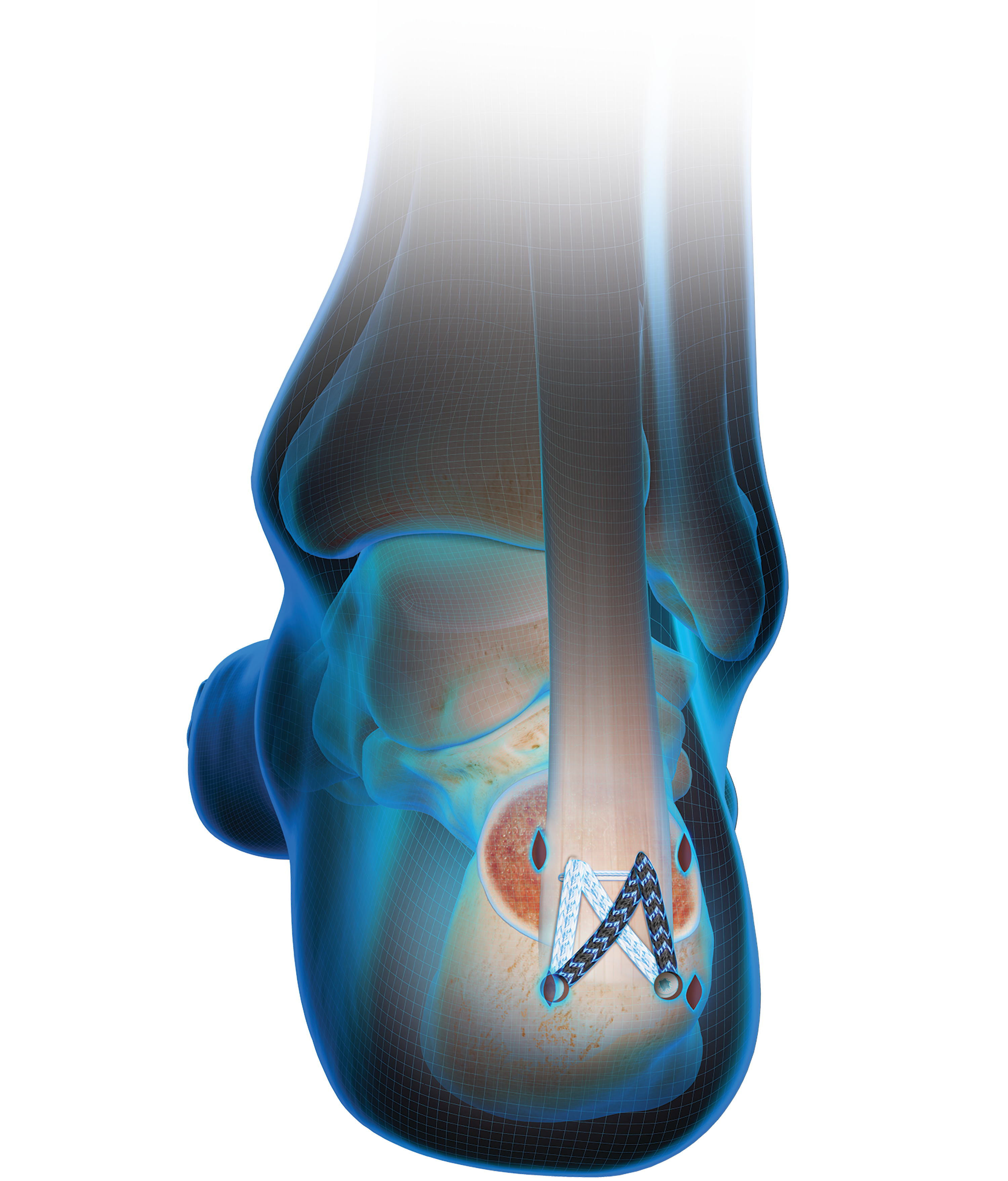
Surgery becomes a consideration when you have a displaced or complicated fracture involving multiple bone pieces or if the injury affects the stability of your shoulder joint. Your surgeon might employ plates, screws, or anchors to secure the bones in their proper place, enabling the restoration of shoulder anatomy and function. After surgery, a period of immobilization is necessary to support the healing process.

Following the initial treatment phase, rehabilitation plays a pivotal role in your recovery. A tailored physical therapy program focuses on gradually restoring strength and range of motion to your shoulder. Persistence and dedication to your rehabilitation exercises will be critical, as they significantly impact your ability to return to normal activities and prevent future shoulder issues.

4 Glenoid Fracture Descriptions

As you navigate the complexities of shoulder joint injuries, knowing the specific type of fracture you’re dealing with can propel your understanding and streamline your path to recovery.

Glenoid fractures are categorized by their location and the pattern of the break within the bone, each type presenting unique challenges and treatment avenues.

Your orthopedic specialist may identify any multitude of these fractures such as Impaction Fractures, where the bone is driven into another, and the more straightforward classifications of Displaced and Non-displaced Fractures, denoting bone alignment post-injury.

Grasping these terms enhances your dialogue with healthcare providers and empowers your active role in the treatment and healing process.

Comminuted Fracture:

A comminuted fracture presents as one of the more serious challenges among the types of glenoid fractures. In your case, this means the glenoid bone is fragmented into three or more pieces, complicating both stabilization and the healing process.

Your treatment may become complex, often involving surgical intervention to reconstruct the fractured bone elements. Restoring the original structure of your shoulder joint becomes a priority to prevent long-term mobility issues and maintain shoulder function.

Impaction Fracture:

Should you face an impaction fracture, it implies a bone has been driven into another, often due to compression or a fall onto an outstretched hand. This type of glenoid fracture sometimes doesn’t present with visually obvious deformity but can cause significant pain and functional impairment in your shoulder.

Your orthopedic care provider will scrutinize this injury meticulously, gauging its impact on your shoulder’s anatomy. It’s not uncommon for treatment to involve a period of immobilization, allowing the impacted bone to settle back into position and fostering recovery with minimal invasiveness.

Displaced Fracture:

Encountering a displaced fracture implies that the bone fragments have been forced out of their original alignment, posing a notable hurdle in your journey to rehabilitation. This condition often arises from high-energy impacts or falls that not only fracture the glenoid but also disrupt the intricate articulation of the shoulder joint.

Addressing a displaced fracture swiftly is essential, as improper alignment can lead to impaired shoulder mechanics and chronic joint issues. Your orthopedic surgeon’s skilled hands will likely intervene, using advanced techniques to realign the bone pieces and stabilize them, ensuring the foundation for your shoulder’s successful recovery is set in place.

Non-displaced Fracture:

A non-displaced fracture means that, despite a break in the glenoid bone, the pieces remain in correct alignment, offering a less complex road to healing. You can breathe a little easier knowing that this type often heals with conservative treatment, bypassing the need for surgical intervention to reposition bone fragments nerve.

Your focus with a non-displaced fracture revolves around allowing your body the necessary time to undertake its natural healing process. Rest and a strategic period of immobilization will likely form the cornerstone of your recovery, paired with careful monitoring by your healthcare provider to ensure the fracture heals appropriately.

Conclusion

Glenoid fractures disrupt the shoulder’s intricate balance, necessitating immediate attention and precise care to restore functionality. Recognizing the signs and symptoms of these fractures enables timely diagnosis and averts potential long-term joint complications.

A variety of treatment methods, including conservative care or surgery, are tailored to the specific nature of the fracture, underscoring the need for specialized medical evaluation.

Ultimately, understanding glenoid fractures empowers individuals to engage actively in their recovery process and to safeguard their shoulder health effectively.


                                                                        (Photo Credit – Shutterstock/Envato/Adobe)

How to Deal With Muscle Knots: Tips for Pain Relief

Shoulder and Lower Pain and Tension Relief - Orthopedic Treatment

Shoulder and Lower Pain and Tension Relief - Orthopedic Treatment

When you feel a stubborn ache in your neck or shoulder, the culprit might be a muscle knot.

These pesky, tight spots of muscle fibers can cause significant discomfort and even headaches if left unmanaged.

But relief is within your reach; with the right approach, you can alleviate the pain and prevent future occurrences.

Armed with the knowledge of effective heat therapy, specialized massage techniques, and stretching exercises, you’ll be on your way to smoother muscles and a more relaxed body.

Keep reading to unlock the mystery of muscle knots and equip yourself with strategies for tackling them head-on.

Understanding the Basics of Muscle Knots

You might be familiar with the sudden pinch or the nagging ache in your muscles that doesn’t seem to relent.

These discomforts are often due to muscle knots, areas where muscle fibers have tensed up and refused to let go, leading to what feels like small, hard lumps within your muscle.

As you tense up, perhaps from stress or overexertion, these fibers contract and create these knots.

Understanding what muscle knots are and recognizing how they form is paramount for effective management.

While they have a tendency to form across various parts of your body, there are certain areas that seem to be more susceptible to knotting, often those you use most frequently or strain the most.

Identifying these common regions can help preempt discomfort, setting the stage for you to learn how to get rid of muscle knots to reclaim comfort and mobility.

What Are Muscle Knots and How Do They Form?

Imagine your muscle fibers as diligent workers on an assembly line, normally operating in harmonious coordination. A muscle knot, or myofascial trigger point, appears when some of these workers go into a state of perpetual overdrive, tensing up into a tiny, localized spasm, stubbornly persisting and interrupting the natural ebb and flow of muscular relaxation.

This unyielding tension disrupts circulation, leading to a build-up of metabolic waste that can exacerbate the discomfort. Your own habits, such as maintaining a single posture for too long or overloading muscles during exercise, often serve as the catalyst for these painful interferences in muscle function.

The Common Areas Prone to Knotting and Why

In your journey to relief, take note of the muscle groups that frequently succumb to knots. Shoulders, neck, lower back, and calves often bear the brunt of these unwelcome guests, largely due to the burden of repetitive movements and the commonality of poor ergonomics in daily activities.

Remember, your posture during prolonged sitting or the repetitive strain from activities like typing or manual labor can predispose these areas to tension. As these muscles carry the responsibility of supporting and moving your body, they fall victim to knotting when overwhelmed or used incorrectly.

Implementing Heat Therapy for Soothing Relief

Turning to heat therapy can be an effective way to alleviate the discomfort caused by muscle knots.

By applying a heat source directly to the affected area, you encourage blood vessels to expand, which enhances blood flow and helps flush away the buildup of waste products contributing to the pain.

Before reaching for the nearest heat pad, though, familiarize yourself with the optimal methods for heat application.

It’s essential to identify the appropriate heat source suited for your specific situation and understand the safety measures to prevent skin damage.

The following discussion will guide you through selecting the ideal heat option and provide you with best practices for applying heat to your muscle knots, ensuring you find relief without risking further injury.

Choosing the Right Type of Heat Source

When selecting a heat source for muscle knot relief, you need to consider the temperature and duration of application that’s best for your body. A heat pad or warm, damp towel might suffice; the key is ensuring the warmth penetrates deeply without causing burns, something that often accompanies excessively high temperatures or prolonged exposure improperly monitored.

Your choice could also involve a localized heat wrap or a warm bath, designed to target broader muscle groups. Seek a solution that provides consistent, therapeutic warmth while accommodating your mobility, as some treatments may be better suited for use during periods of rest or activity.

Best Practices for Applying Heat to Muscle Knots Safely

When applying heat to muscle knots, it’s paramount that you gauge the heat, ensuring it’s warm enough to relax the muscles but not so hot that it scalds your skin. Opt for a heat setting that allows you to apply it for longer periods, typically around 15 to 20 minutes, which can significantly aid in reducing the muscle tension without causing discomfort or damage to your skin.

Always use a barrier, such as a cloth or towel, between your skin and the heat source to protect against burns. Evenly distributing the warmth across the affected muscle area promotes efficient circulation and aids the healing process. Consistent checks of skin temperature and color during the heat application will help you maintain a safe environment for your muscle healing journey.

Exploring Massage Techniques at Home

Turning your attention to the transformative power of touch, exploring massage techniques can be a pivotal strategy for relieving muscle knots.

The right approach to massaging your muscles can be akin to conducting a well-orchestrated symphony where every movement works towards soothing those tight, knotted areas.

Self-massage tools, if used correctly, can amplify your efforts, providing targeted relief with precision.

From foam rollers to therapeutic massage balls, these instruments, when accompanied by the proper technique, can yield promising results.

Equally important is mastering the technique of hand massages, which enables you to apply direct pressure to affected areas, bringing about immediate respite.

Up next, you’ll discover how to wield these tools effectively and navigate through a step-by-step guide to hand massaging tight muscles, paving your path to greater comfort and reduced pain.

Self-Massage Tools and How to Use Them Effectively

Unlocking the potential of self-massage tools begins with choosing the right instrument for your needs. A foam roller, for instance, can serve as an invaluable ally, particularly for larger muscle groups; to use it effectively, position yourself so the affected area experiences gentle, sustained pressure to encourage release and relief.

On the other hand, maneuvering a massage ball requires a different strategy, one that involves small, deliberate movements to target those deep-seated knots. Apply moderate pressure using the ball on the tense area, moving slowly to stimulate blood flow and relax the tight fibers without overwhelming your muscles.

Step-by-Step Guide to Hand Massaging Tight Muscles

Initiate the process of hand massaging by gently placing your fingers over the affected muscle knot. Use a firm, but not crushing, grip to apply steady pressure with slow, intentional strokes that follow the grain of the muscle fibers. This methodical approach can coax the rebellious contractions into smoother territories, easing away tension and fostering recovery.

Transition your touch into deeper circular movements as the muscle begins to yield. Your intent should be to communicate with the knotted fibers, nudging towards relaxation without eliciting pain. This tactile conversation encourages the proper distribution of blood flow, leading wayward muscle fibers back to their natural rhythm of tension and release.

Incorporating Stretching Exercises Into Your Routine

Embarking on a quest to unravel the discomfort of muscle knots often leads to the therapeutic world of stretching.

Synonymous with relief, stretching exercises should take on a pivotal role in both your pain management and prevention tactics.

They underpin the continued flexibility and strength of your muscles, fostering an environment where knots struggle to maintain their grasp.

By introducing gentle stretches specifically designed to target the muscle groups prone to knotting, you channel much-needed attention to areas asking for relief. Furthermore, stretching adjacent areas that lead to muscle imbalance can also provide relief and prevent further muscle knots from arising.

Establishing a daily routine focused on stretching not only addresses current soreness but successfully builds your muscular defense against future tension.

Going forward, let’s explore how to effectively integrate these stretches into your everyday life for an enduring reprieve from the shackles of muscle knots.

Gentle Stretches for Targeted Muscle Relief

Embarking on gentle stretches translates to sending a signal of ease to your tense muscles, particularly those afflicted by the stubbornness of muscle knots. When you stretch, do so with intention and mindfulness, focusing on breath and alignment to ensure the stretch caters to alleviating tension and promoting muscular harmony. These stretches are designed to be kind to your body, allowing the muscle fibers to elongate and release in a gradual and controlled manner.

Adopt a routine that prioritizes stretches targeting your neck, shoulders, and back, areas frequently gripped by knots due to everyday demands. As you guide these parts of your body through the stretches, maintain a pace that is slow and attentive, granting each muscle group the time it needs to respond to the relief being offered, laying the groundwork for a lasting respite from discomfort.

Creating a Daily Stretching Schedule for Prevention

You know the importance of regular exercise, and the same principle applies when preventing muscle knots. Establish a stretching schedule that aligns with your daily life, ensuring that it’s practical and sustainable for long-term adherence.

Initiate and end your day with a series of stretches designed to target key areas susceptible to tightness. Your consistent dedication to this routine will play a significant role in maintaining muscle health and preventing the formation of new knots.

The Role of Hydration and Nutrition in Preventing Muscle Knots

Adequate hydration is vital to your muscle health, as water plays a critical role in maintaining normal muscle function and healing. Ensuring you drink ample fluids throughout the day keeps muscle tissues supple and less prone to the development of painful knots.

Beyond quenching your thirst, your dietary choices have a direct impact on muscle recovery and tension prevention. Incorporating foods rich in magnesium, such as almonds and spinach, can aid in muscle relaxation and mitigate the risk of creating tight spots within your muscle fibers.

Lean proteins, packed with essential amino acids, serve as the building blocks for repairing and strengthening muscle tissue. Including these in your meals helps fortify your muscles against the unnecessary stress that often leads to knot formation.

By nourishing your body wisely and maintaining hydration, you lay the foundation for resilient muscles that are less susceptible to the discomfort of muscle knots. Thoughtful attention to diet and water intake can be a transformative approach to managing and preventing those tenacious knots.

Conclusion

Addressing muscle knots requires a proactive combination of targeted heat therapy, massage techniques, and regular stretching to alleviate pain and improve muscle function.

Implementing these strategies into your routine can greatly enhance blood flow, relieve tension, and foster muscle recovery.

Maintaining proper hydration and nutrition bolsters your body’s defenses against the formation of new knots, keeping muscles supple and resilient.

Carefully curated, these pain relief methods intertwine to form a comprehensive approach to managing and mitigating the discomfort of muscle knots.


                                                                        Photo Credit – Adobe

Jacksonville Orthopedic Surgeon Earns Award for Invention to Repair Achilles Tendon

Jorge Acevedo Headshot

Dr. Jorge Acevedo wins prestigious Edison Award, recognized for innovative implant and technique

JACKSONVILLE, FL (June 19, 2024) – Not too many doctors can say they have invented something to help their patients. However, Dr. Jorge Acevedo, a nationally recognized Foot and Ankle surgery expert with Southeast Orthopedic Specialists, cannot only say he helped invent an important advance in orthopedic care, but also won an award for it.


Recently, the Jacksonville orthopedic surgeon earned the prestigious Edison Award – often referred to as the “Oscars of Innovation.” Just like the Olympics, winners in various categories are given a gold, silver, or bronze award. Dr. Acevedo and New York-based Dr. James McWilliam won the gold award, recognized for developing the Arthrex MIS FiberTak Achilles SpeedBridge Repair Implant System. This innovative system is used to repair the Achilles tendon, the large tendon at the back of the heel.


“We are proud to support Dr. Acevedo and Dr. McWilliam in their quest to revolutionize Achilles tendon repair,” said a spokesperson for Arthrex, Inc. “Their innovative implant system exemplifies our commitment to empowering surgeons with cutting-edge technology that improves patient outcomes.”


The Achilles tendon is comprised of strong tissues that connect the calf muscle to the heel. In some instances, the tendon may get damaged through overuse, physical activity or repeated stress and may require surgery.


With traditional Achilles tendon surgery, surgeons may need to open the back of the heel to repair the tendon. There may be large incisions, potential damage to surrounding tissue, major scarring and recovery time could be extensive.


Dr. Acevedo’s invention involves a minimally invasive procedure and specialized technique that minimizes damage to surrounding tissue. A series of innovative instruments and implants are used. Small incisions are made in the back of the heel and strong sutures are placed to hold the tendon against the heel bone.

MIS Achilles Blue Wire Illustration

As the area heals, patients are left with minimal scarring and a faster recovery time compared to traditional surgery.

“We are honored to receive the Thomas Edison Gold Award for our work in advancing orthopedic care,” said Dr. Acevedo. “Our goal was to develop a solution that not only effectively addresses Achilles tendon pathology but also prioritizes patient comfort and recovery.”


For more information, please visit www.se-ortho.com. Click “here” to view an animation of the Arthrex system.


About Southeast Orthopedic Specialists

Established in 2001, Southeast Orthopedic Specialists is a regional leader in musculoskeletal care. Southeast Orthopedic Specialists was founded on the principle of providing high-quality, comprehensive care in the Northeast Florida region. The entire spectrum of orthopedic care is provided to patients, including orthopedic surgery, spine care, physiatry, pain management, chiropractic, podiatry and physical therapy. Physicians at Southeast Orthopedic Specialists are leading experts in their field with decades of experience, including the completion of multiple fellowships and groundbreaking research. Visit www.se-ortho.com to learn more.


About Healthcare Outcomes Performance Company (HOPCo)

Healthcare Outcomes Performance Company (HOPCo) is the national leader of integrated musculoskeletal value-based health outcomes management, practice management and health system service line management. This includes comprehensive expertise across all MSK specialties, including orthopedics, spine, neurosurgery, hand, pain management, rehabilitation and neurology. HOPCo’s integrated care, analytics, and facilities development and management platforms have proven to increase the quality of patient care while reliably reducing the total cost of care across the care continuum for practices, health systems and payors alike. HOPCo’s affiliated payors, practices, and health systems successfully participate in highly efficient value-based contracting (bundled payments, chronic episodes of care initiatives, population health programs, and other advanced risk-based arrangements) utilizing HOPCo’s proprietary platforms, IT solutions, integrated analytics, and evidence-based comprehensive care pathways. Visit https://hopco.com to learn more.


For media inquiries, please contact:


David Lozano
Senior Director of Communications
Healthcare Outcomes Performance Company (HOPCo)
18444 N. 25th Ave.
Phoenix, AZ 85023
623.241.8779
David.lozano@hopco.com

Close
Close

Schedule an Appointment

Loading...
Get the Care You Need, When You Need It.

You now have the ability to schedule an appointment online any time with our orthopedic surgeons and specialists. Our intelligent system connects you to the right doctor in the right setting at the right time. Schedule your appointment online above!
*Providers will be recommended based on specialty and best availability. If you would prefer to schedule an appointment with a provider of your choice, please call us at 904-634-0640